Again, no major theme or single lesson this week. Just continuing to throw a few random ideas out.
And back to i-pads and apps and Cell Ultrastucture. TEM exercise updated and scanned June 2014I’ve always done it like this – even if they just need information, it’s better for them to be reading and processing and using the information, rather than just copying it down. But this exercise has been transformed by the i-cell app
which allows them to not only see all the structures in helpfully colour coded 3D, but to spin them, manipulate them, zoom in on them…
I quickly follow this with “Snot Trafficking” Protein trafficking new for 2014 Sept which I link to the splendid John Kyrk animation, so that they can see how all the organelles interact. A clip from Secret Life of Cell showing Kinesin in action on the cytoskeleton illustrates how transport vesicles are moved (and brings in the role of the Mitochondria). These superb clips of vesicles moving in real time is another vivid punctuation mark in their activity.
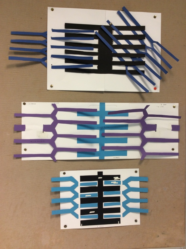
They will find this difficult. This year I livened it up by turning the lab into a cell. Starting in one corner with the festive DNA model
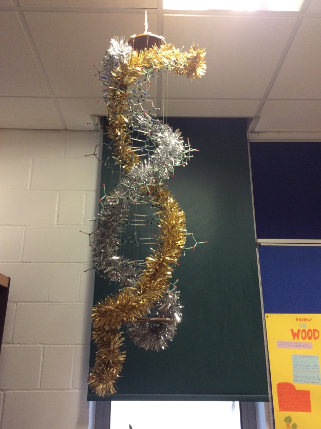
I modelled the copying of the message (just a note on a post it – green, red, blue, red, green, yellow, blue etc) passed on to a student playing a ribosome with a large pile of coloured chunky lego (amino acids) which she had to join together in the order specified.
The finished lego polypeptide is put into a tray (a transport vesicle) and moves to the next desk (the first layer of the Golgi). On the desk, more students add post-its (carbohydrate chains) before putting it into another “vesicle” and shuffled on to the next desk. I made loud squelching noises to illustrate the budding and fusing of the vesicles from one desk to the next.
The final desk is linked to the Fire Exit by a bit of string – and I walk the tray along it to the door which I then open and throw the lego out of the lab with a flourish (they really liked this bit).
The resulting A3 posters were superb as they threw themselves into the detail…
And Year 13 and muscles.
As a springboard into micro-fibrils, try giving them an Electron Micrograph of muscle tissue (unlabelled) and ask them to draw it. They will produce a beautiful observational drawing of a sarcomere without having any idea of what it is. Ask them to describe it. How could this work? They’re half way towards the sliding filament model which, once they’ve figured it out, they can then make a working model of ….